 Culturing Life: How Cells Became Technology by Hannah Landecker is an extensive history of the culturing of cells in the lab. As such, it gave many details about the fits and starts involved in the early attempts to get cells to grow reliably. As is usually the case in scientific research, as much is owed to timing and serendipity as to careful repetition and fastidious lab work.
The idea that cells could be taken from an organism and cultured separately was initially met with skepticism. The general thinking was that cells could not become autonomous from the organism. By 1885, Wilhelm Roux showed that he could keep nerve cells from chicken embryos alive for several days in the lab. Once the principal was established, scientists began to search for the right media, the right glassware, and the right cells or tissue to make cell culture reliable.
 The growth in virology was a major driver for the improvement of cell culture. Virologists needed a way to make large-scale cultures of infected samples for vaccine development. Early vaccines were grown using embryonated chicken eggs, a method that had a high yield for virus, but was quite costly. (Flu vaccines are still made using chicken eggs; the specifics of the process are described here.) By the 1930s, the yellow fever vaccine was the first to be produced with cultured cells, but it was not clear that these approaches would work for other viruses.
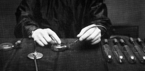 |
| Alexis Carrel used specially designed flasks for growing cells |
It is important to consider Enders' work in the context of time and place. As Landecker writes, "It is not that Enders was particularly good at growing human cells." Instead, like so many scientific breakthroughs, Enders' success was due to being in the right place at the right time with the right reagents. The location of his lab in Boston Children's Hospital afforded Enders a ready supply of living tissues (from abortions, miscarriages, hysterectomies, and circumcisions, which were all used without concerns about patient consent or privacy). The timing was also critical as cell culture was becoming more feasible due to improved techniques and the increased availability of antibiotics. Enders' methods became the basis for the production of virus cultures for vaccine development, most significantly the creation of polio vaccine by Jonas Salk in 1954.

 The next major event in the history of cell culture happened in 1951, when an African-American patient named Henrietta Lacks was treated for cervical cancer at Johns Hopkins. Lacks' biopsy came into the hands of George Gey, who was able to generate the first human cell line (called HeLa cells) that in culture continuously. HeLa cells are unique in many respects: they grow rapidly and are robust enough to withstand shipping and freeze/thaw
cycles. These unique features (which have been explained to some degree by the genome sequence) were what allowed researchers to culture the cells so
easily, making HeLa cells a standard cell line in most laboratories.
The next major event in the history of cell culture happened in 1951, when an African-American patient named Henrietta Lacks was treated for cervical cancer at Johns Hopkins. Lacks' biopsy came into the hands of George Gey, who was able to generate the first human cell line (called HeLa cells) that in culture continuously. HeLa cells are unique in many respects: they grow rapidly and are robust enough to withstand shipping and freeze/thaw
cycles. These unique features (which have been explained to some degree by the genome sequence) were what allowed researchers to culture the cells so
easily, making HeLa cells a standard cell line in most laboratories.The story of the origins of the cell line that was generated from Ms. Lacks' biopsy has been told in beautiful detail by Rebecca Skloot in The Immortal Life of Henrietta Lacks. Skloot's book (which is on my list of top science reads) focuses on the Lacks family as it comes to terms with the Henrietta's legacy. In Culturing Life, Landecker tells the HeLa story in broader strokes with a different historical context, focusing on how the tenor of the HeLa cell line origin story has changed over time. In 1968, with tissue culture techniques established and many cell lines available, Stanley Gartler published a Nature paper profiling eighteen cell lines; using the presence of a gene variant only present in African Americans, he showed that all were contaminated by HeLa cells. Predictably, these results created a major stir in the research community. Landecker details the language used to describe HeLa cells, particularly in regards to the contamination issue: aggressive, surreptitious, and malicious. Some scientists suggested that "one drop was enough" to contaminate and ruin a culture, which is evocative of the one-drop rule of racial classification in the US. Thus, unlike for most cells, the race and gender of the donor was central to the discussion of the cells.
While the language used to talk about HeLa cells has changed considerably, some elements have remained consistent. Scientists and science writers still connect the cells with Henrietta Lacks and talk about how the cells have allowed her to achieve immortality. Most articles will also detail how many HeLa cells have been grown since Gey started to culture the cells. Indeed, these are fascinating details. According to Skloot's book, "One scientist estimates that if you could pile all HeLa cells ever grown onto a scale, they’d weigh more than 50 million metric tons—an inconceivable number, given that an individual cell weighs almost nothing. Another scientist calculated that if you could lay all HeLa cells ever grown end-to-end, they’d wrap around the Earth at least three times, spanning more than 350 million feet." Thus, while the legacy of HeLa cells may be complicated, their utility in the research lab is not.
