Thursday, March 31, 2016

How my PhD training has improved my baking skills

I walked away from the bench four years ago this month. Since that time, I have enjoyed the life of a Scientific Editor, where my training in the lab is integral to the job. I am still surprised when my training becomes useful in everyday life. Perhaps it shouldn't –  many articles about "The PhD Problem" focus on how training to be a scientist has other long-term benefits, including developing critical thinking and problem solving skills. A few years ago, I decided that I would start approaching recipe development like I would designing a protocol or experiment. This has been a very fun project, so I wanted to share my results.

First, I started reading more about recipe development by professional bakers. Then, I decided to tackle my favorite baked good: the oatmeal cookie. For many years, I have used my mom's recipe, which came from the Quaker Oats box and was never altered. They are delicious cookies, but as any scientist/baker would, I wondered if I could do better.


I had to figure out what recipe to start with. The Flour cookbook has a great oatmeal cookie recipe, with excellent instructions that are thoughtful about the science of baking (e.g., the formation of gluten). Another advantage is that their recipe includes weights for all the ingredients. The use of a scale to weigh ingredients was a crucial element for my recipe testing – it added precision and allowed me to make half a recipe (this meant I could bake cookies every other weekend without having too many extra calories). The Flour cookies are very good, but I found them too chewy and I wanted more oatmeal. I did some more research on how each ingredient in a cookie recipe contributes to the end result. (The best resource was The Food Lab.) Over the course of a year, I baked four different iterations of the Flour recipe and ended up with something in between their recipe and the original Quaker recipe. I have repeated this protocol six times and find it is consistently good – even after we moved (testing a new oven) and got new cookie sheets. Out of curiosity, I did try America's Test Kitchen recipe (free version here), as ATK is the gold standard of recipe testing. I found their cookie had a good oat flavor, but was too crunchy and crumbly. 
This experiment taught me a lot about recipe design and the small things that make a big difference in how cookies turn out (e.g., letting the dough rest overnight). I also found many useful resources to help me in future recipe development. It is definitely a great time to be a scientist/baker as there are so many food blogs and podcasts that focus on this topic. The success of my experiment and the wealth of resources available has encouraged me to approach my previously perfected "Test Kitchen Brownies". I will be sure to update you once my next experiment is complete.

Thursday, February 11, 2016

DIY Science: Biopunk by Marcus Wohlsen shows how to do science away from the bench

I have read many stories about doing science away from the bench, both in the news and views section of scientific journals and on popular science blogs. PLOS Blogs even has a regular feature called Citizen Sci, which highlights how everyday people can take part in science outside the traditional laboratory or field setting. Thus, I was curious to read Biopunk: DIY Scientists Hack the Software of Life by Marcus Wohlsen.

Wohlsen did a great job of discussing what DIY science is and why it is important. Historically, the roots of DIY science can be found in people like Edward Jenner, who inoculated his gardener's son with cowpox to test its efficacy against smallpox and Newton, who poked himself in the eye to better understand optics. Today, the DIY science movement serves to democratize science, allowing people outside of research institutes and universities to take on projects based on their own interests rather than based on the decisions of funding bodies. As a result, the movement could transform science, taking some power away from the big companies that set steep prices for lab equipment.

One major goal of the DIY science movement is to MacGyer common lab equipment to make it accessible (read: cheap) to everyone. Some of these hacks could serve researchers that aren't lucky enough be in a well-funded lab. Take the example of the PCR machine, the biology lab workhorse that helps researchers copy and manipulate DNA (prices start at $3000). A Google search for "DIY PCR machines" reveals many designs that start at $100, making PCR available to more people. Thus, many of the projects in DIY biology can benefit the developing world because they are design to cut costs and to make it possible to run experiments in virtually any environment.

One element that seems to be common in every story about DIY biology is the comparison with the computer hacking movement. People turning spare rooms and garages into laboratories for their side projects does have similarities with the origin stories of companies like HP and Apple. The resemblance goes further: DNA is the code of life and, like computer hackers, people in the DIY bio movement believe that this code should be open source and not limited only to the scientists who have grant money or research labs. Labeling DIY scientists as "hackers" can create a negative impression; when the general public hears of a hacker doing genetic modification in their garage, there can be a tendency to overreact. The worst case was seen with Steve Kurtz, an artist that used the tools of DIY biology as part of his palette. In 2004, Kurtz was arrested on suspicion of bio-terrorism after his wife died of an unrelated heart attack. (There is also an interesting documentary about the case called Strange Culture.)

Wohlsen pushes the idea that DIY scientists are punks, describing their leather jackets, tattoos, and Mohawks, to the point that borders on fetishization of his interview subjects. To some extent, this is SOP in science writing (and celebrity interviews); it serves as a device to quickly characterize the subject. Unfortunately, it detracts from the serious science that is starting to happen. I look forward to seeing what DIY bio leads to in the future, especially because CRISPR technology has made genome editing simpler and cheaper than ever and it is readily available to DIY biologists.

Another great aspect of DIY science is that it can give the people who left the lab after their PhD or postdoc a way to return to the excitement of scientific discovery. Depending on where you live, you may find a hacker space near you. Here in Boston, you can visit the BossLab (Boston Open Source Science lab), but most major cities seem to have similar spaces available. Some more great resources can be found at DIYbio.org. Of course, I think I will keep my home experiments confined to baking. 

Monday, November 30, 2015

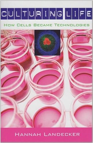
Culturing Life reveals the history of tissue culture, with some interesting details about HeLa cells

Culturing Life: How Cells Became Technology by Hannah Landecker is an extensive history of the culturing of cells in the lab. As such, it gave many details about the fits and starts involved in the early attempts to get cells to grow reliably. As is usually the case in scientific research, as much is owed to timing and serendipity as to careful repetition and fastidious lab work.

The idea that cells could be taken from an organism and cultured separately was initially met with skepticism. The general thinking was that cells could not become autonomous from the organism. By 1885, Wilhelm Roux showed that he could keep nerve cells from chicken embryos alive for several days in the lab. Once the principal was established, scientists began to search for the right media, the right glassware, and the right cells or tissue to make cell culture reliable.

The growth in virology was a major driver for the improvement of cell culture. Virologists needed a way to make large-scale cultures of infected samples for vaccine development. Early vaccines were grown using embryonated chicken eggs, a method that had a high yield for virus, but was quite costly. (Flu vaccines are still made using chicken eggs; the specifics of the process are described here.) By the 1930s, the yellow fever vaccine was the first to be produced with cultured cells, but it was not clear that these approaches would work for other viruses.

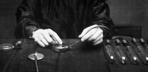
Alexis Carrel used specially designed flasks for growing cells
In the late 1940s, John Enders started using cell culture to grow viruses. His lab was the first to show that polio virus could be cultured in human tissues and that infection with the virus caused rapid changes in the appearance of the cells; these results meant that there was a quick and reliable assay for infection. Enders published his results in the journal Science in 1949 and soon after received the Nobel prize for his work.

It is important to consider Enders' work in the context of time and place. As Landecker writes, "It is not that Enders was particularly good at growing human cells." Instead, like so many scientific breakthroughs, Enders' success was due to being in the right place at the right time with the right reagents. The location of his lab in Boston Children's Hospital afforded Enders a ready supply of living tissues (from abortions, miscarriages, hysterectomies, and circumcisions, which were all used without concerns about patient consent or privacy). The timing was also critical as cell culture was becoming more feasible due to improved techniques and the increased availability of antibiotics. Enders' methods became the basis for the production of virus cultures for vaccine development, most significantly the creation of polio vaccine by Jonas Salk in 1954.

Importantly, Enders and Salk also had a major role in organizing the tissue culture community. One major push was for standardization of reagents and media; in the early days of cell culture, each lab made its own glassware and media, so it was nearly impossible to share cell lines between labs. Another proponent of standardization was cell biologist and electron microscopist Keith Porter. Porter wanted to image whole cells by EM, but was frustrated by the need to learn the exacting and often finicky methods of cell culture to get his experiments done. Porter and others started a group that would later become the Tissue Culture Association, which aimed to standardize media preparation and other elements of tissue culture.


The next major event in the history of cell culture happened in 1951, when an African-American patient named Henrietta Lacks was treated for cervical cancer at Johns Hopkins. Lacks' biopsy came into the hands of George Gey, who was able to generate the first human cell line (called HeLa cells) that in culture continuously. HeLa cells are unique in many respects: they grow rapidly and are robust enough to withstand shipping and freeze/thaw cycles. These unique features (which have been explained to some degree by the genome sequence) were what allowed researchers to culture the cells so easily, making HeLa cells a standard cell line in most laboratories.

The story of the origins of the cell line that was generated from Ms. Lacks' biopsy has been told in beautiful detail by Rebecca Skloot in The Immortal Life of Henrietta Lacks. Skloot's book (which is on my list of top science reads) focuses on the Lacks family as it comes to terms with the Henrietta's legacy. In Culturing Life, Landecker tells the HeLa story in broader strokes with a different historical context, focusing on how the tenor of the HeLa cell line origin story has changed over time. In 1968, with tissue culture techniques established and many cell lines available, Stanley Gartler published a Nature paper profiling eighteen cell lines; using the presence of a gene variant only present in African Americans, he showed that all were contaminated by HeLa cells. Predictably, these results created a major stir in the research community. Landecker details the language used to describe HeLa cells, particularly in regards to the contamination issue: aggressive, surreptitious, and malicious. Some scientists suggested that "one drop was enough" to contaminate and ruin a culture, which is evocative of the one-drop rule of racial classification in the US. Thus, unlike for most cells, the race and gender of the donor was central to the discussion of the cells.

While the language used to talk about HeLa cells has changed considerably, some elements have remained consistent. Scientists and science writers still connect the cells with Henrietta Lacks and talk about how the cells have allowed her to achieve immortality. Most articles will also detail how many HeLa cells have been grown since Gey started to culture the cells. Indeed, these are fascinating details. According to Skloot's book, "One scientist estimates that if you could pile all HeLa cells ever grown onto a scale, they’d weigh more than 50 million metric tons—an inconceivable number, given that an individual cell weighs almost nothing. Another scientist calculated that if you could lay all HeLa cells ever grown end-to-end, they’d wrap around the Earth at least three times, spanning more than 350 million feet." Thus, while the legacy of HeLa cells may be complicated, their utility in the research lab is not.

Wednesday, October 7, 2015

Frankenstein's Cat by Emily Anthes looks at how genetic engineering is changing the animals around us


Frankenstein's Cat: Cuddling up to Biotech's Brave New Beasts by Emily Anthes explores how biotechnology and genetic engineering are changing the animals around us. The book delves into the science behind these new creatures as well as the ethical issues and public perceptions. The result is an easy and interesting read that makes you imagine the genetically engineered animals that could be on the horizon.


GloFish were the first commercially available pets created using biotechnology. By inserting the gene that makes jellyfish glow (green fluorescent protein, or GFP) into a common pet store find (zebrafish), the inventor was able to create a fish that glowed green in certain lights. Despite seeming relatively benign, the company was required to perform extensive research on the possible environmental impacts in case of escape before GloFish were approved for sale in 2003. Interestingly, consumers had few concerns about these new creatures and were willing to pay almost 20 dollars for each fish. The company now has a variety of other colors and species available and sells special tanks to help you enjoy their fluorescent fish.

Pharming is a branch of biotechnology where researchers use farm animals to produce a range of pharmacological products. For example, goats can be engineered to produce the protein lysozyme in their milk. Lysozyme has been shown to inhibit bacterial growth; some preliminary results suggest that the lysozyme-enriched milk can improve the immune system. The scientists behind this project hope that this goat milk could help protect children from bacterial infections. Similar approaches have been used to engineer goats that have silk protein in their milk and silkworms that make collagen instead of silk. These additives are not harmful to the animals and have a range of potential applications.

The chapter on animal cloning starts with Dolly the sheep (the first cloned animal) and makes some interesting stops along the way. The story of Carbon Copy (CC), the first cloned cat (sometimes called Copy Cat), is scientifically notable because CC doesn't look much like her calico mother Rainbow. This is due to a process called X chromosome inactivation (you can learn more about this process in my recent post on Junk DNA). The gene for orange fur is on the X chromosome. CC's genome has an inactive copy of the X chromosome from her clone mother. Thus, while CC is genetically identical, she is phenotypically different. This result was interesting to scientists, but it likely gave future customers of cat cloning some pause–what's the point of paying to have your favorite cat cloned if you can't be sure you will get a cat that looks the same?

Anthes tours a wildlife preserve and research center in Louisiana where researchers aim to develop and perfect methods for cloning animals with the long-term goal of preservation of endangered species. This is certainly worthwhile goal, but the barriers, both technical and ecological, are numerous. To me, the biggest problem with saving a species through cloning is the lack of genetic diversity. Until these problems are solved, many are banking on frozen zoos, large stocks of samples from a variety of animals that are endanger of extinction. The hope is that once cloning technology improves, it may become viable to add to the existing populations.

Roboroach from Backyard Brains
The chapter on cyborg animals was truly fascinating. For example, Anthes talks about the CIA's experiments with remote-controlled cats. Project Acoustic Kitty was predictably a failure due to the fiercely independent nature of cats. There are some notable success stories with insect cyborgs. In fact, for 99 dollars, you can build your own Roboroach. The newest approaches include the use of optogenetics, where specific neurons are made to be light sensitive, allowing researchers to control animals by shining a light. These experiments have some important applications, such as the removal of land mines and the detection of survivors in earthquake rubble, but people find animal mind control to be unsettling (even if it is a roach). It is important to consider that.

The book concludes with predictions of what may be possible in the future, like creating farm animals that are resistant to disease to decrease the use of antibiotics. Frankenstein's Cat was published in 2013 and the field has made amazing progress since that time. With the advent of the new genome editing technique CRISPR, the possibilities are truly endless, which has led to concerns about the ethical and safety considerations of this technology. Earlier this week a Chinese Institute caused a stir with its announcement of the commercial availability of gene-edited micro-pigs as pets. It will be interesting to see where we draw the line on genetically modified animals.  

Wednesday, September 23, 2015

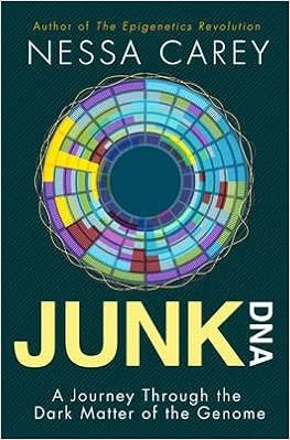
Junk DNA: Nessa Carey's new book about the actually important stuff in the genome.

In 2001, when the first draft of the human genome was completed, researchers were surprised to learn that only 2% of the human genome codes for proteins. At the time, scientists were very focused on proteins and thought that there would be a much larger number of protein-coding genes in the human genome due to our complexity. The term "junk DNA" has been used to describe the other 98% of the genome. With only 20,000 protein-coding genes, the human genome contains almost the same number of genes as the simple roundworm and model organism C. elegans. However, C. elegans has very little excess DNA, suggesting that this junk DNA could be part of the explanation for the increased complexity of humans. This is the starting point for Nessa Carey's second book Junk DNA: A Journey Through The Dark Matter of the Genome, which explains the importance of the non-coding portion of the genome.

Some scientists have argued that the term junk DNA should be scrapped for a more neutral term like non-coding DNA. They suggest that the term is dated and inaccurate. In addition, calling it junk is rather pejorative and is based on the protein-focused view of the genome. Carey's book nicely demonstrates that the other 98% isn't always junk.

What is the other 98% of the genome good for then? Some non-coding DNA has well-established functions. For example, the centromeres are the stretches of DNA that allow the chromosomes to attach to the cell's chromosome segregation apparatus (the mitotic spindle) when the cell copies and divides its DNA. Another example is the telomeres, the lengthy repeat regions of DNA at the ends of the chromosomes. Because telomeres shorten with every cell division, they are linked with aging.


Junk DNA also encodes several special types of RNAs, including long non-coding RNA (lncRNA), microRNA (miRNA), and small interfering RNA (siRNA), that control gene expression. One of the earliest described examples of these special RNAs is found in the biology of sex determination. In XX females, one X chromosome is inactivated to ensure that genes on the X chromosome are not overexpressed. This process, called X chromosome inactivation, is controlled by a gene called Xist (X-inactive specific transcript). Xist encodes a long non-coding RNA, which covers one X chromosome and inactivates it (Xi). Interestingly, on the opposite strand from Xist is a gene called Tsix, which is expressed on the active X chromosome (Xa). The expression of these genes is mutually exclusive, ensuring that only one X chromosome is activated. The Xist/Tsix story highlights the power of special RNAs in controlling gene expression. These RNAs are the subject of intense research in both basic and clinical settings. Carey describes several approved drugs and promising clinical trials based on anti-sense approaches.

In short, Junk DNA was quite readable and should be informative for readers at any level of knowledge about molecular biology. My only complaint about the book was Carey's decision not to include protein or gene names in her writing. In the first chapter, she explains that this was due to the fact that half of her readers find it disruptive. Instead, where applicable, she includes footnotes with the gene or protein names. Unfortunately for me, I am in the half that finds it disruptive to read footnotes to learn the name of the gene in question. Otherwise, the book was very up to date and comprehensive. I also liked her use of simple graphics to explain complex concepts in molecular biology. I recommend Junk DNA for those who want to learn more about why the non-coding regions of our DNA are not junk.

Sunday, September 13, 2015

The Emperor of All Maladies - comments on the second part of the PBS special

In all my reading about cancer biology, I have not yet tackled The Emperor of All Maladies, which is said to be the best book on the subject.  Luckily, PBS and Ken Burns have delivered an excellent three-part series based on Siddhartha Mukherjee's 2011 book. This post covers the contents of part two, "The Blind Men and The Elephant."

This part of the series focuses on discovering the cause of cancer. The title, an allusion to the parable, refers to the fact that for many years scientists could not find the connection between the three major causes of cancer: viral, chemical, and genetic. The ideas were separated ideologically and scientifically. At conferences, the scientists that supported each of these ideas did not interact. It was only relatively recently that the connections between these causes were illuminated.

The earliest carcinogen was discovered in 1911 by Peyton Rous, who described the viral origin of avian sarcoma (for more information check out this great story by Jessica Wapner). In 1964, Burkitt lymphoma was linked to the Epstein-Barr virus. These results led to an rapid increase in the focus on viral carcinogenesis with the idea that a vaccine could prevent cancer. This focus came at the cost of other ideas about the causes of cancer. Unfortunately, Human papillomavirus (HPV) and Hepatitis (HepB and C) have been the only other viral carcinogens identified.

The second idea was that chemicals cause cancer. Lung cancers became increasingly common in the late 1940s. Epidemiological studies showed links between cigarette smoking and lung cancer, but tobacco companies obfuscated the results. In 1964, scientific links between cigarettes and lung cancer were firmly established thanks in part to the Kennedy administration's blue ribbon panel tasked with investigating the matter. Once the epidemiological methods were established for tobacco, other chemicals were added to the carcinogen list.

The final idea was that genes caused cancer. The major breakthrough came from Michael Bishop and Harold Varmos, who were studying the Rous sarcoma virus. Their timing was perfect – the tools of molecular biology were becoming readily available. Work from their labs led to the discovery of a gene called Src, the first described oncogene. The oncogene idea was that normal genes in our bodies that control cell growth can be turned on at high levels and cause cancer. Robert Weinberg later identified the first human oncogene, Ras. Dozens of other oncogenes were found in subsequent years, leading to optimism that the cure for cancer was surely close at hand. However, we have since learned that cancer is a complex disease (some argue a collection of diseases) with a diverse range of etiologies, which makes it impossible to treat with a one-size-fits-all approach.

Interspersed with the description of cancer research was a narrative of one woman's treatment for breast cancer. This story begins with a history of breast cancer treatment, including a discussion of William Halsted's radical mastectomy. As I covered in more detail in my recent post on Pandora's DNA, Halsted's method was the standard treatment for breast cancer for nearly a century. The success rate was not impressive, but the treatment approach was unchallenged until Bernard Fisher criticized its use. Fisher performed a clinical trial to compare the use of the lumpectomy with the radical mastectomy. In 1985, his results showed that either approach was just as effective, but that the lumpectomy was less invasive and led to improved quality of life. Radical mastectomy was no longer the standard therapy: "cutting more did not mean curing more".

The intention of this segment was to illustrate the personal side of cancer, but for me the link between the two segments was how the treatment of cancer has evolved in parallel with developments in cancer research. This highlights how basic scientific research is a critical starting point for successful clinical outcomes.

Tuesday, August 18, 2015

The genetic wonders of red hair

I recently finished reading Armand Leroi's Mutants, where I learned about some of the polymorphisms (small changes in DNA sequence) linked to variations in human skin and hair color, particularly red hair. With two different types of redheads in the house, I have always been curious about the genetic basis of this relatively rare trait.


red-haired mouse (from Flickr)
Hair and skin color are determined by the relative proportions of the two types of melanin pigment: eumelanin (dark brown) and pheomelanin (red/yellow). Large amounts eumelanin result in darker hair and very little produces blond hair. People with more pheomelanin have red hair. Red hair phenotypes can range from pale red to bright red or reddish brown, which is due to a balance of the two melanin types. Polymorphisms in the melanocortin 1 receptor gene (MC1R) are associated with variations in hair and skin color in mammals. The MC1R protein is a membrane receptor found only in melanin-producing cells (melanocytes). In response to melanocyte-stimulating hormones (MSHs), MC1R initiates a cascade of cellular events that turns on the production of pigment synthesizing genes, including pheomelanin or eumelanin. Essentially, MC1R determines pigmentation by regulating the relative proportion of eumelanin and pheomelanin.


The ancestral form of the MC1R allele produces eumelanin; variant alleles are less functional, decreasing eumelanin production or increasing the amount of yellow/red melanin. Variations in MC1R contribute to a spectrum of phenotypes, including freckling, red hair color, and sun sensitivity. MC1R alleles that disrupt function are present in ~80% of individuals with red hair and ~20% with brown-black hair. More than 80 MC1R allele variations have been identified in European populations. Association studies have shown that certain MC1R variants (p.D84E, p.R151C, p.R160W, p.D294H, p.R142H, and p.I155T) are linked with the red hair color phenotype.

One unanswered question is why these polymorphisms are more prevalent in Eurasians than in Africans. The current hypothesis is that paler skin permits better synthesis of Vitamin D in environments with less sunlight. A recent study using data from the 1000 Genomes Project revealed that the polymorphisms are also more common in Northern Europe than Southern Europe, which supports the Vitamin D hypothesis. Interestingly, a MC1R variant has been observed in Neanderthals, indicating that red hair and pale skin were also present in this population. A recent paper has shown that the Neanderthal MC1R variant is rare in Europeans, but can be found in East Asian populations. This result suggests that mutation of MC1R was a common mechanism to adapt to changes in sunlight intensity.

from David Fisher's lab


Some MC1R variants are associated with an increased risk of melanoma. Due to increased sun sensitivity and freckling of people with pale skin, this relationship seems obvious. However, darkly pigmented Caucasians with certain MC1R variants also show an increased incidence of melanoma. This result suggests that the MC1R pathway may have another role in the development of melanoma beyond differences in the ability to filter UV rays in light and dark skin. In fact, scientists think that MC1R may play a pigment-dependent and a pigment-independent role in skin carcinogenesis. There are a few hypotheses to explain the link between MC1R and melanoma. First, after UV exposure, cells with more pheomelanin show increases in DNA damage, which is correlated with increases in abnormal cell growth and proliferation, a hallmark of cancer. Second, a recent paper in Molecular Cell suggested that UV light triggers the interaction of a tumor suppressor called PTEN with MC1R. The tumor suppressor can interact with wild type MC1R, but not the red hair alleles of MC1R. The PTEN-MC1R interaction protects PTEN from degradation, which suppresses an oncogenic signaling pathway (PI3K/Akt). In contrast, MC1R variants do not interact with PTEN, allowing increased levels of oncogenic signaling pathways after UV irradiation. Unfortunately for redheads, sun exposure alone is not the sole mechanism for skin cancer. A Nature paper from David Fisher’s lab used a red-head mouse model with inactive MC1R to investigate a possible UV-independent pathway. They found that in the presence of the most common melanoma oncoprotein (BRAF 600E), ginger mice developed melanoma without UV exposure, while MC1R wild-type mice did not. Thus, shielding easily freckled skin from the sun may not be enough to protect from skin cancer for people with certain MC1R variants.


The association between red hair and melanoma suggested that there may be a scientific basis for the headline from 2014 that climate change was putting the red hair gene in danger of extinction. The story was exposed as alarmist and scientifically inaccurate. One of the many problems with the article is that they label the red hair gene as recessive. Because hair color is a complex phenotype, it is not surprising that the genetics of red hair are also complex. Red hair is usually inherited in a recessive manner, but it can also be dominant. A 2000 study showed that the inheritance pattern depends on the MC1R allele that is present: most alleles are recessive, but some alleles can be dominant. Individuals that are heterozygous for a mutant MC1R allele show variations in red hair color, beard color, or freckling. Thus, there is a dosage effect of MC1R variants on hair and skin color, which explains why some men have red beards and brown hair. These results also explain how two red-haired parents can (on rare occasions) have children that don't have red hair. Thankfully, the many variations in MC1R and the complex inheritance patterns mean that red hair isn't likely to die out any time soon.

**************************

For more information, I recommend the following links:

Red heads feel pain differently A blog post from 23andme about the population genetics of red hair.